Things we can see in your eyes.

As part of every eye check we examine your eyes with a microscope - have you ever wondered what we can see? Here is the big reveal!

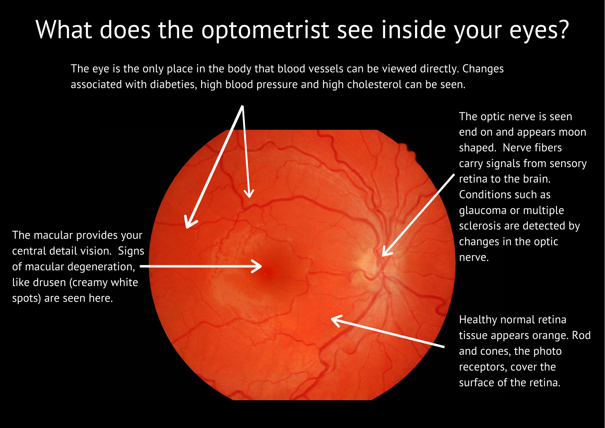
What does the optometrist see inside your eyes_.png

A lot of the tissue in the eye is transparent - so that light can pass through to get to the retina. Let’s start at the front - the cornea. We often see evidence of previous infections or injuries. In the fluid filled chamber inside the front of the eye our microscope allows us to see cells floating. Checking the lens we look for early signs of cataract. Then we see the vitreous gel, where we often find the ‘floaters’ which people describe for us. Finally the retina comes into focus.

The retina is red - sometimes seen as a red eye in photos taken using a flash. We have a check list as we examine the blood vessels, the optic nerve and the macula.

Sometimes we want to go deeper - to ‘look’ at the layers of the retina in more detail. This is when we use OCT scanning technology. While the information this provides is quite amazing, it does not quite match to viewing down the microscope - seeing individual red blood cells tumbling along the capillaries at the front of the eye will always be special for me.

What is an OCT? How does it help in Macular Degeneration and Glaucoma?

OCT stands for scanning technology Optical Coherence Tomography. Scanning lets us ‘see’ tissue behind the visible retina inside the eye. In the past 5 years OCT has become invaluable in assessing eye health, particularly helpful for Macular Degeneration (MD) and Glaucoma.

Copy of Copy of poster OCT.jpg

When we see signs of MD during an eye examination, we take an OCT scan which reveals what is happening in the underlying retina. In particular we want to spot the ‘wet’ form of MD which can cause rapid, devastating vision changes. With a scan we can see if there is fluid in the macula and allows us to make referral for treatment.

The detailed measurements of the nerve fibre layer in the eye are particularly helpful when we are looking for Glaucoma. If there are signs in a regular examination, OCT gives us a way to closely monitor changes over time. These changes are seen before any vision loss.

But OCT is not a stand alone magic bullet. It’s a tool and interpreting the results needs careful consideration along with all other clinical findings. At the end of the day an OCT scanner is not an optometrist.

We are committed to using all our available diagnostic tools and we strongly believe the best place to start is regular eye examinations. The simple, vital step of spending time with you, to collect relevant history, through careful listening, guides us to the best combination of diagnostic testing.