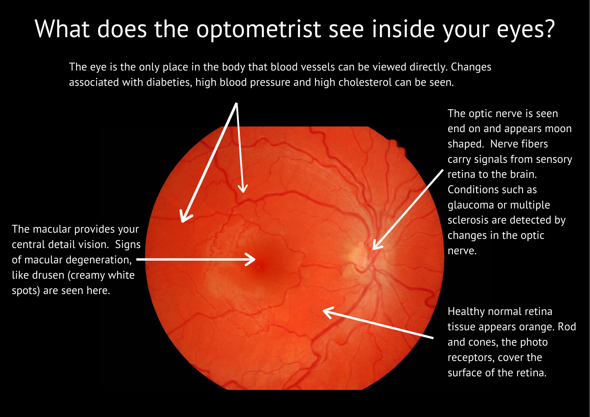
A lot of the tissue in the eye is transparent - so that light can pass through to get to the retina. Let’s start at the front - the cornea. We often see evidence of previous infections or injuries. In the fluid filled chamber inside the front of the eye our microscope allows us to see cells floating. Checking the lens we look for early signs of cataract. Then we see the vitreous gel, where we often find the ‘floaters’ which people describe for us. Finally the retina comes into focus.
The retina is red - sometimes seen as a red eye in photos taken using a flash. We have a check list as we examine the blood vessels, the optic nerve and the macula.
Sometimes we want to go deeper - to ‘look’ at the layers of the retina in more detail. This is when we use OCT scanning technology. While the information this provides is quite amazing, it does not quite match to viewing down the microscope - seeing individual red blood cells tumbling along the capillaries at the front of the eye will always be special for me.


